
Page 21 - COS-FOD2015
P. 21
Compend. Oral Sci:vol1(3);2015;14-20
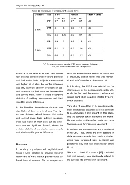
Table 2: Mandibular interradicular distance (mm)
Cut level Site Male Female t-test P value
Mean SD Mean SD
P-P 3.7 0.8 3.7 0.7 NS
P-M 4.1 0.9 3.8 1.2 NS
6 mm
M-M 4.4 0.85 4.6 0.7 NS
P-P 3.9 0.8 3.5 1.2 NS
P-M 4.2 1.5 4.2 1.4 NS
8 mm
M-M 4.6 1.1 4.2 1.2 NS
P-P, first premolar-second premolar; P-M, second premolar- first molar;
M-M, first molar- second molar. NS, not significant
higher at 8 mm level in all sites. The highest jects had normal vertical relation as this is also
root distance existed between second premolar a previously studied factor that was demon-
and first molar. Male subjects’ measurement strated to influence bone dimensions [16].
was higher at all sites, but gender difference
In this study, the C.E.J was selected as the
was only significant at 8 mm level between sec-
starting point for the measurements, unlike oth-
ond premolar and first molar and between first
er studies that used the alveolar crest as a ref-
and second molar. Table 1, shows descriptive
erence point, which could be affected by perio-
statistics of maxillary measurements and t-test
dontal problems.
result for gender differences.
Yang et al [3] stated that in the anterior maxilla,
In the Mandible, interradicular distance was
most interradicular distances were not sufficient
also higher at 8 mm level in all sites. The high-
to accommodate a mini-implant. In this study,
est root distance existed between first molar
only the posterior part of the maxilla and mandi-
and second molar. Male subjects’ measure-
ble was studied as they offer a wider and more
ment was higher at most sites, but the differ-
favourable area for miniscrew placement.
ence was not significant. Table 2, shows de-
scriptive statistics of mandibular measurements In addition, our measurements were conducted
and t-test result for gender differences. using CBCT files, which are more accurate in
distance measurements than previous studies,
which were conducted using periapical and
Discussion panoramic x-ray that have magnification errors
[9, 10].
In our study, only subjects with sagittal skeletal
Class I were included as previous research Min et al [11]and Kuroda et al [12] concluded
shows that different skeletal pattern shows dif- that root proximity was significantly related to
ferent bone dimensions. Also all sample sub- the success rate of miniscrew placement .
17